
Avinent apostamos por la felicidad común ofreciendo soluciones dentales que cambian la vida de las personas
 Implantes dentales
 Implantes dentales
 Las soluciones implantológicas de Avinent se han situado en la vanguardia de la implantología digital. La fabricación de implantes dentales es nuestro producto estrella y están diseñados con la total seguridad de contar con una superficie patentada y probada en todo el mundo. Además, nuestro sistema de cirugía guiada encaja a la perfección con todo el ecosistema para utilizarse de una forma sencilla y eficaz. Esto nos permite conseguir obtener resultados predecibles y excelentes.
Centro de fresado y sinterizado
 Centro de fresado y sinterizado
 El flujo digital de Avinent supone una evolución sin precedentes en los procesos de elaboración protésica, tanto en diseño como en desarrollo y fabricación de estructuras. Por consiguiente, en Avinent utilizamos la última tecnología en torneado, fresado e impresión 3D para ofrecer un universo de soluciones. Como resultado, el fresado de estructuras de alta precisión es uno de los productos mejor valorados del sector dental con una calidad reconocida internacionalmente.
Componentes protésicos
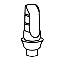 Componentes protésicos
 Para dar una solución completa y cerrar un círculo de alto valor añadido, en Avinent producimos componentes protésicos, tanto digitales como tradicionales, para implantes de todas las marcas. Por lo tanto, en Avinent contamos con un flujo digital establecido y una calidad excelente en aditamentos y componentes protésicos para llevar a cabo de principio a fin los tratamientos implantológicos. De hecho, en la Tienda Online de Avinent, puedes acceder al amplio catálogo de soluciones en un clic.
Craneomaxilofacial
 Craneomaxilofacial
 Como resultado de la utilización de la impresión 3D como base del proceso productivo, y la experiencia adquirida a través de la innovación tecnológica, en Avinent ofrecemos soluciones en el campo craneomaxilofacial (CMF). Una solución digital completa que ofrece a hospitales y clínicas, implantes personalizados, guías quirúrgicas, cirugías reconstructivas, cirugías de ortognática, ATM y otras cirugías de la zona facial. En consiguiente, en Avinent también ofrecemos la impresión de modelos 3D para facilitar a los centros médicos el estudio de los casos más complejos.
Ortodoncia
 Ortodoncia
 Fruto del trabajo en I+D+I, y de la capacidad productiva en el ámbito de la impresión 3D y de los materiales, en Avinent lanzamos alineadores invisibles personalizados para resolver todos los retos estéticos y funcionales. Recode Aligners, es la marca de alineadores de Avinent, que además de tratar multitud de casos con éxito, ofrece todos los recursos de comunicación para que la clínica pueda llegar a los pacientes. De igual manera, nuestra línea también incorpora otras soluciones ortodónticas como los retenedores y las férulas de descarga.